

LASER stands for Light Amplification by Stimulated Emission of Radiation. It is a high energy beam of light which is used to treat the eyes in various ways. LASERs used in the OPD (Out-patient department) are mainly used for the management of retinal and anterior segment conditions.
Retinal LASER
These are done usually to the swelling in the central part of the retina (Macular oedema), for areas of ischaemia in retina especially when abnormal new vessels are formed or to weak areas in retina (holes, tears).
Procedure and preparation
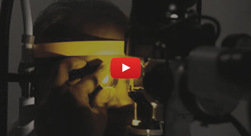
The eyes will need to be dilated prior to the treatment. LASER can be delivered either by indirect ophthalmoscope (LIO) or by slit lamp. LIO is usually preferred if the area to be treated is in the far periphery or when the media is very hazy due to cataract or vitreous haemorrhage. A contact lens needs to be applied on the eye before slit lamp delivery of LASER. Hence, anaesthetic drops are applied before commencing slit lamp delivery of LASER. The procedure usually lasts for about 5-15 minutes.
Red LASER (810 nm)
Due to its larger wavelength, the LASER penetrates deeper tissues. It also penetrates better in eyes with retinal haemorrhage. It is usually used for treating the abnormal retina in babies with Retinopathy of prematurity (ROP)
Green LASER (532nm)
This is the conventional form of retinal laser and is commonly used when the media is clear. This LASER can be delivered by LIO or by slit lamp. The hospital can deliver this laser through a slit lamp as a single spot or as multiple spots.
Yellow LASER (577nm)
A yellow laser (577 nm) is used for selected macular conditions, including diabetic macular oedema, central serous chorioretinopathy and age-related macular degeneration. This wavelength scatters less light and can pass through some media opacities, such as cataract and vitreous haemorrhage. Compared with a conventional green laser, its effect is less dependent on melanin in the retinal pigment epithelium and more dependent on haemoglobin in the choriocapillaris. It is minimally absorbed by macular pigments (xanthophylls) and well absorbed by oxygenated haemoglobin. It is also used for micropulse subthreshold laser therapy in diabetic macular oedema, branch retinal vein occlusion and central serous retinopathy. With this mode, exposures as low as 10 milliseconds are possible, so treatment can be given without a visible tissue endpoint. The hospital can deliver this laser through a slit lamp as a single spot or as multiple spots. Suitability is decided after a clinical examination.
Disadvantages of conventional LASER
More pain as exposure time cannot be less than 100 ms
Worsening of macular oedema. Multiple sessions of conventional LASER are advocated in order to overcome this disadvantage Expansion of spot size resulting in scarring and RPE atrophy.
Visual field defects and reduced dark adaptation
The above disadvantages result in a poorer patient experience thereby adversely affecting their compliance to the therapy
Multiple spot LASER
A multispot pattern laser photocoagulator can deliver several laser spots to the retina in a preset pattern. The exposure time is shorter than with conventional laser, which can reduce discomfort. The spots are uniform. Spot expansion, retinal pigment epithelium atrophy and scarring are limited, so collateral tissue effect is smaller. Inflammation is also lower, which can reduce laser-induced macular oedema. Whether this form of laser is suitable depends on the examination.
Indications for retinal lasers
- Diabetic Macular oedema
- Proliferative diabetic retinopathy
- Macular oedema due to retinal vein occlusions
- Neovascularisation secondary to retinal vein occlusions
- Peripheral retinal degenerations/holes/tears
- Eales disease and other retinal vasculitis
- Central serous retinopathy
- Retinopathy of prematurity
- Extrafoveal polyps of polypoidal choroidal vasculopathy (PCV)
Rarely
- Coat’s disease
- Familial exudative vitreo retinopathy
- Retinal Artery Macroaneurysm
- Ocular ischaemic syndrome with secondary neovascularisation


Anterior segment LASER
Nd-YAG LASER (1064 nm) is used for anterior segment laser.
This is usually of 2 types i.e capsulotomy and iridotomy.
Capsulotomy
This is done for posterior capsular opacification Which is opacification of the posterior capsule days to yeas after cataract surgery. The opacification results in reduced vision. Posterior capsulotomy makes an opening in the capsule resulting in improved transmission of light into the eye thereby enhancing the vision. The procedure is painless. Eye drops are advised for 1 week after the procedure after which the vision is reassessed to look for improvement & dispensing spectacles

Iridotomy
This is done to relieve pupillary block or in eyes with narrow angles which might result in increased intraocular pressure causing optic nerve damage (Glaucoma).
In this procedure, drops are applied to constrict the pupil. After constriction is adequate, anaesthetic drops are applied and a contact lens is used to visualise the area to be treated. An alternate pathway is thus created to facilitate the drainiage of excess fluid tapped behind the iris. The procedure is painless and drops are advised for a week after the procedure. After a week the intraocular pressure and angles are reassessed by the ophthalmologist.