



Advanced 3D Surgical Technology
NGENUITY 3D visualisation for digitally assisted vitrectomy and selected vitreoretinal surgery.
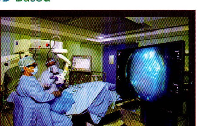
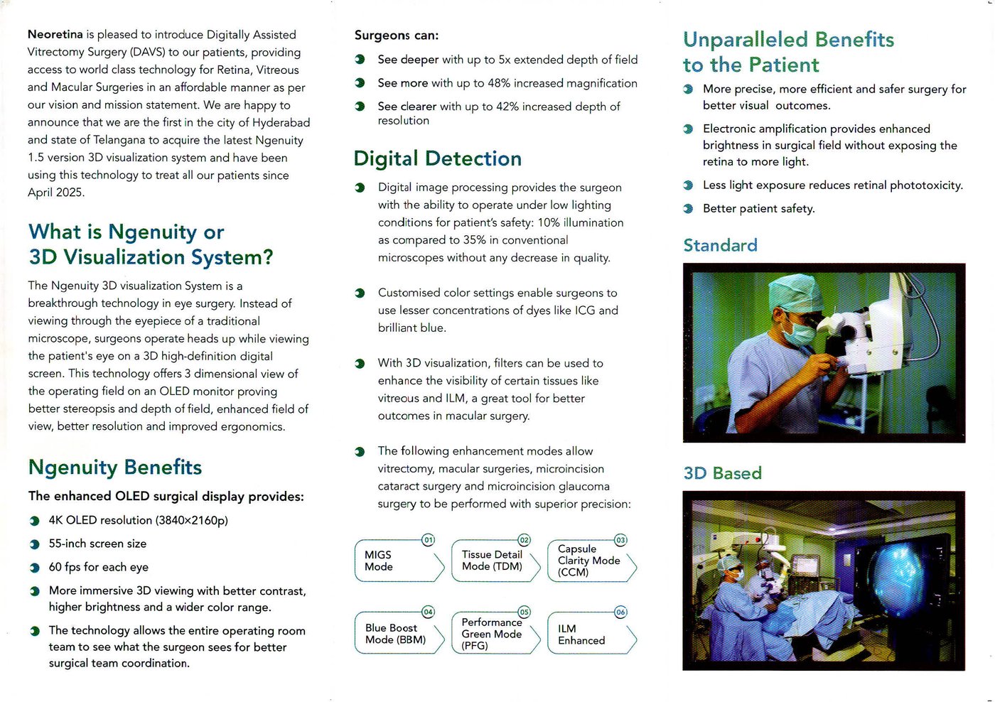
Neoretina uses the NGENUITY 1.5 3D visualisation system for digitally assisted vitrectomy surgery (DAVS) and selected retina, vitreous and macular procedures. Since April 2025 it has been available at Neoretina as a first in Hyderabad and Telangana.
Heads-up 3D surgery
Instead of looking only through microscope eyepieces, the surgeon works from a high-definition 3D OLED display. The operating microscope image is shown on a large screen so the surgeon and the theatre team share the same view of the surgical field.
What the system provides
- 4K OLED display (3840 × 2160) on a 55-inch screen, with a stereoscopic view for the operating team.
- Digital control of brightness, magnification and colour during the operation.
- Extended depth of field and higher magnification compared with a conventional microscope view, as described by the manufacturer.
- Used for selected vitreoretinal surgery alongside the operating microscope; the choice depends on the diagnosis and the surgeon's assessment.
Digital visualisation modes
Colour and contrast settings can be adjusted to highlight structures such as vitreous and the internal limiting membrane (ILM). Modes available on the system include Tissue Detail, Capsule Clarity, Blue Boost, Performance Green and ILM Enhanced, among others.
Lower microscope illumination can be used while the display amplifies the image electronically. That can reduce light reaching the retina during longer procedures. Whether a particular mode is used depends on the case.
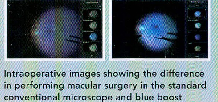
How this may help patients
- A clearer, shared 3D view can support precise work in complex retina and macular surgery.
- Electronic image amplification can keep the field bright while limiting light exposure to the retina.
- Colour settings may allow lower concentrations of surgical dyes when staining membranes, according to clinical need.
Outcomes still depend on the eye condition, the procedure and recovery. The system is one part of surgical planning; it does not replace clinical judgement.
Ready when you are
Book an appointment
Speak with our team on WhatsApp, or visit us in Hyderabad.
